8902235337
Guide to Laparoscopic Myomectomy – How It Is Done, Benefits, Risks and More
September 30, 2025
Across the world, millions of women suffer from uterine fibroids. One of the most common gynecological problems worldwide, it is non-cancerous in form. Although benign, these can lead to many discomforting symptoms. Women can suffer from bloating, pain in their pelvis or even bleed heavily during their menstrual cycles. They can also experience fertility problems.
Naturally, it is important to diagnose this problem accurately so that it can be managed or treated effectively. For a long time, doctors used 2D ultrasound for diagnosis. Now, 3D ultrasound or 3D USG for fibroids has come up. It has brought a revolution in the way the diagnosis and assessment of uterine fibroids are carried out.
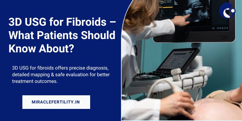
It is an advanced imaging technique that offers detailed 3D images of internal organs, such as the uterus. Standard 2D imaging scans offer a single-plane, flat image. 3D ultrasound, however, compiles multiple images obtained from various angles to create a volumetric data set. Doctors can use this to view the fibroids and uterus in greater detail. They can view these from multiple perspectives and in cross-sections. Such types of images are impossible with 2D scans.
For women who are suffering from fibroids, a 3D ultrasound can be very useful. It can offer a comprehensive and precise view of the fibroids, including their exact location, number, and size, as well as how they are connected to the uterine cavity. With this crucial information, doctors can diagnose and plan treatment more conveniently.
The 3D ultrasound process starts similarly to a traditional ultrasound. A probe, generally of a transvaginal nature, emits audio waves that bounce off the uterine tissues. The returning echoes are recorded in the form of sequential 2D images. The variance lies in the fact that 3D ultrasound grabs multiple images in various planes – axial, horizontal, and perpendicular. It then makes use of a specialized software program to compile them into a 3D volume.
When this 3D volume data is created, doctors can manipulate and review it from different angles. They can get multiplanar views, which can offer better clarity than regular scans. It is possible to visualize fibroids in relation to the uterine cavity, walls and nearby structures in a more accurate way.
The biggest advantage is that 3D ultrasound can offer a detailed, holistic view of the uterus, which is impossible with traditional scans. With the coronal and multiplanar views, especially, doctors can conduct a more comprehensive evaluation of fibroids. 3D imaging offers a more comprehensive evaluation than 2D scans, which may sometimes end up missing deeply embedded or small fibroids.
Due to this improved visualization, doctors can perform diagnoses more accurately. They can easily differentiate fibroids from adenomyoma, adenomyosis, or other gynecological conditions.
There are plenty of other benefits, such as:
Fibroids can develop in various layers of the uterus, such as on the outer surface, within the wall or inside the cavity. It is important to determine the exact location and type, to decide the right treatment plan. With 3D ultrasound, doctors can locate fibroids and determine their characteristics precisely. They can easily understand the relationship of the fibroid to the uterine cavity and wall.
Fibroids are not the only condition to result in heavy bleeding or uterine enlargement. Adenomyosis, for example, can resemble fibroids in 2D scans. 3D ultrasound, with its multiplier imaging, can help physicians to distinguish between these conditions. There can be more accuracy in diagnosis and treatment can be more appropriate.
3D ultrasound helps to create a detailed map of the uterus, which can be especially useful for women who suffer from multiple fibroids. It can show the position, size and number of each fibroid. Such type of mapping can be especially useful while planning hysteroscopic myomectomy, laparoscopic myomectomy or other surgical interventions.
3D ultrasound can also offer a coronal view, or a cross-sectional view of the uterus, right from the top to the bottom. This is one of its most significant benefits. When physicians get this kind of perspective, they can quickly evaluate the relation of fibroids to the uterine fundus and endometrial lining. This proves to be critical in the assessment of fertility and treatment strategies.
3D ultrasound, just like 2D ultrasound, makes use of soundwaves rather than radiation. Due to this reason, it is safe to be used repeatedly, even in women who are planning to conceive or are pregnant. It is painless, non-invasive and comes with no known risks.
It can be used in these cases:
When physicians consider the use of surgery for removing fibroids, it is essential for them to have accurate knowledge of their size, relation to the uterine cavity and location. With 3D ultrasound, surgeons can get detailed anatomical information that they need for planning minimally invasive processors with more safety and accuracy.
The classification of fibroids is done on the basis of their location such as subserosal (growing outward on the uterine surface), submucosal (protruding into the uterine cavity), and intramural (within the uterine wall). 3D ultrasound can be very effective in the evaluation of all three types of fibroids, including small-sized fibroids that regular scans may end up missing altogether.
Fibroids may sometimes interfere with embryo implantation or distort the uterine cavity in women who are suffering from infertility problems. With the aid of a 3D ultrasound, doctors can easily evaluate how fertility might be impacted by fibroids. They can take proper decisions about whether they should go for surgical removal of fibroids before pursuing IVF or other similar treatment methods.
3D ultrasound has revolutionized the way uterine fibroids are diagnosed and managed. It addresses many of the limitations of traditional ultrasound and can definitely help gynecologists to ensure better care and treatment for patients. In all those cases where there are risks of severe complications, this procedure can be a lifesaver in all possible aspects.

September 30, 2025

September 29, 2025
September 25, 2025